


Frequently Asked Questions about Anencephaly
by Monika Jaquier
- What is anencephaly?
- What role does the brainstem play?
- Occurrence?
- Children most affected?
- Can it be treated?
- Manifestation?
- What causes an anencephaly?
- Is it caused by anything the parents did?
- Life expectancy?
- At what point can an anencephaly be diagnosed?
- What is AFP?
- Is the diagnosis reliable?
- Might the mother's health be jeopardised?
- What does a child with anencephalylook like?
- Can a child with anencephaly sense or do anything?
- What happens during the birth of a child with anencephaly?
- Can children with anencephaly be organ donors?
- What is the rate of recurrence?
- Can anencephaly be prevented?
- What research is being done?
- References
What is anencephaly
Anencephaly is a congenital birth defect (from the Latin congenitus "born with"). It begins to develop right at the
start of life in the womb. The word anencephaly means "without an encephalon", the encephalon being the set of nervous
center contained in the brain. This is not an entirely accurate definition: whilst a child with anencephaly is indeed
born without a scalp, without a vault of the cranium, without meninges, without either brain hemisphere and without a
cerebellum, the child is nevertheless usually born with part of its cerebral trunk, brainstem (Müller 1991). Infants
with anencephaly may still have small foci of histologically normal cerebral cortex (Stumpf et al 1990)
Almost 75% of babies with anencephaly born at term survive their birth. The life expectancy of those who survive is only a few hours or
days (Jaquier 2006).
Approximately 20 percent of affected infants have additional congenital anomalies (Botto 1999).
What role does the brainstem play?
Together with the spinal chord, it controls many of the body's unconscious functions, such as the heart beat, and it
coordinates most voluntary movements.
Occurrence:
Approximately one child for every 1000 births (Central Europe). This rate varies according to populations. (Sadler, T.W. 2005)
Children most affected:
Anencephaly affects more girls than it does boys.
Can it be treated?
Unfortunately, there is no known treatment for a child with an anencephaly.
But this doesn't mean that nothing can be done for the baby and his family. Many hospitals
offer perinatal hospice care. A perinatal hospice approach walks with these families on
their journey through pregnancy, birth, and death, honoring the baby as well as the baby's
family. Perinatal hospice is not a place; it is more a frame of mind. It is a way of caring
for the pregnant mother, the baby, the father, and all involved with dignity and love.
Learn more about perinatal hospice.
Manifestation:
Anencephaly belongs to the family of neural tube defects. A neural tube defect (NTD) is a congenital malformation which
occurs between the 20th and 28th day after conception (Sadler 1998). The cells of the neural plate make up the foetus'
nervous system. In normal development, they fold back onto themselves in order to create what is called the neural tube,
which then becomes the back bone and the spinal cord. After a number of transformations, the superior pole eventually
becomes the brain. One can liken this process to a coin whose edges merge at its centre. In the case of an
NTD, the neural tube is unable to close completely. Anencephaly occurs when the
head end of the neural tube fails to close.
Infants with this disorder are born without a scalp or cerebellum. Their meninges, both hemispheres of the brain and the
vault of the cranium/skull are also missing, though they usually do have part of the brain stem. The remaining brain
tissue is protected only by a thin membrane. About ¼ of babies with anencephaly die before or during birth; those who survive
have a life expectancy of a few hours or days (Jaquier 2006).
What causes an anencephaly?
It is still not known what causes anencephaly. It is probably triggered by a combination of genetic and environmental
factors (Sadler 2005). We nevertheless know that taking Folic Acid can prevent anencephaly. Some medicines (the pill,
valproic acid, antimetabolic drugs and others) lower Folic Acid levels, hence taking them increases the risk of giving
birth to a child with anencephaly (Sadler, 2005).
Ninety-five percent of infants with anencephaly are born in families with no history of neural-tube defect.
Once an infant with anencephaly has been born, the risk increases to 2 to 5 percent in subsequent pregnancies (Stumpf 1990)
Chromosomal abnormalities, single-gene mutations, and teratogenic causes are indentified in fewer than 10 percent of
affected infants (Holmes 1976)
Is it caused by anything the parents did?
No, no one is to blame for the baby having anencephaly.
Life expectancy:
About 7% die during the pregnancy (prenatal death), 20% die during delivery; 50% have a life expectancy
of between a few minutes and 1 day, 23% live more than 1 day (Jaquier 2006).
In very, very few cases, the child can live for weeks or even months.
Get more information: Report about the birth and life of babies with anencephaly
At what point can an anencephaly be diagnosed?
Anencephaly can be reliably diagnosed at 11-14 weeks of gestation by ultrasound scan (Johnson SP et al. 1997).
With the advances in ultrasound-technology, diagnosis tend be possible even earlier. Fleurke-Rozema (2015) found that factors
determining successful early diagnosis are competence level of the sonographers, with a significantly higher detection rate when scans were performed
by a sonographer licensed for nuchal translucency measurement and gestational age at or beyond 11 weeks of gestation.
AFP levels can be measured via a maternal serum screening test (blood test).
If levels are high, there is a risk that the child may be suffering from an NTD.
Further tests must then be carried out (ultrasound scan or amniocentesis) to determine whether there really is a
problem.
What is AFP?
The foetus, through its urine, releases into the amniotic liquid a protein called alpha-fetoprotein (AFP). The exposed
tissue of a child suffering from an NTD release greater quantities of AFP into
the amniotic liquid. The AFP then enters the mother's blood stream through the
placenta and can hence be measured.
Is the diagnosis reliable?
Anencephaly is a malformation which is very easy to see on an ultrasound scan. If a qualified specialist has made an
ultrasound scan diagnosis after the 11th week, the likelihood of a misdiagnosis is minimal.
A positive maternal serum screening test, however, simply shows that there is a higher risk that the baby has Trisomy
21 or 18, or an neural tube defect. Most women who test positive give birth to healthy babies. Additional tests must
be carried out to determine whether the baby is suffering from one of those ailments.
Might the mother's health be jeopardised?
No. Pregnancy is unaffected. In around a quarter of cases, too much amniotic liquid is produced (polyhydramnios). This
is due to the fact that the child does not have the reflexes to enable it to swallow the amniotic liquid. If the volume
of liquid is excessive, it can cause discomfort for the mother. Labour may be triggered prematurely, or waters may
break. An amniocentesis can then be carried out to reduce the amount of liquid (amniodrainage); Amniotic liquid is removed with a
syringe, thus providing the mother with temporary relief.
What does a child with anencephaly look like?
The body of a child with anencephaly is entirely unaffected. However, the vault of the cranium is missing from the eye-brows up. Half-way up to the back of the head is usually covered by skin and hair. Vivid dark red neural tissue covered by nothing more than a thin membrane can be seen through an opening in the head. The size of this opening varies considerably from one child to another. The eyeballs can protrude because of a malformation of the eye-sockets, which is why children with anencephaly are sometimes pejoratively described as looking like frogs.
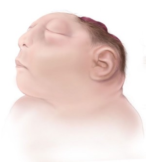
Illustration used with the permission of CDC Centers for Disease Control and Prevention
Can a child with anencephaly sense or do anything?
Doctors will tell you that a child with anencehaly can neither see nor hear, nor feel pain, that he or she is a vegetable.
However, that does not match up with the experience of many families who have had a child with anencephaly. The brain is
affected to varying degrees, according to the child; the brain tissue can reach different stages of development. Some
children are able to swallow, eat, cry, hear, feel vibrations (loud sounds), react to touch and even to light. But most
of all, they respond to our love: you don't need a complete brain to give and receive love – all you need is a heart!
What happens during the birth of a child with anencephaly?
Normally, the baby helps to trigger labour with its pituitary gland and suprarenals (glands of the kidneys). However,
these are either missing or their development has been stunted in children with anencephaly, hence labour does not always
begin spontaneously. As a result, many women ask that labour be induced at the end of their pregnancy. As the vault of
the cranium is missing, it is important that the waters do not break for as long as possible during labour so that they
can exert the necessary pressure on the cervix for it to dilate. If it is possible to keep the waters intact, the birth
of a child with anencephaly will happen in almost the same way as if the mother were giving birth to a healthy child, and
will take as long. The experience of mothers of children with anencephaly has shown that the artificial breaking of waters
significantly reduces the chances of the baby being born alive (Jaquier 2006).
Can children with anencephaly be organ donors?
Since 2012, donation possibilities for babies, specifically babies with anencephaly,
have expanded and currently there are more potential for donation than ever before
in the United States. Although each particular case is unique, and each potential
donation has various criteria that must be met, donation is a very viable option for
a baby with anencephaly if families wish to pursue this option.
There are two main types of donation possibilities: Donation for transplant and donation for research.
Learn more about the possiblity of donation and find links on our special page.
What is the rate of recurrence?
In most cases it is an isolated anomaly and it is very unlikely that it should occur again in the same family.
Statistically, the rate of recurrence for a woman who has already had a child with anencephaly is 4%.
Can anencephaly be prevented?
For some time now, the aetiology of Neural Tube Defects has cited diet and environmental factors. Clinical studies have
confirmed that taking a vitamin called Folic Acid reduces the risk of developing a Neural Tube Defect. If all women of
child-bearing age took 0.4 mgs of Folic Acid every day before conception and at least until the end of the first term of
pregnancy, 50 to 70% of potential cases of anencephaly and Spina Bifida could be prevented (Ceizel and Dudas, 1992).
Learn more about the prevention of neural tube defects.
What research is being done?
The National Institute of Neurological Disorders and Stroke conducts and supports a wide range of studies that explore
the complex mechanisms of normal brain development. The knowledge gained from these fundamental studies provides the
foundation for understanding how this process can go awry and, thus, offers hope for new means to treat and prevent
congenital brain disorders including neural tube defects such as anencephaly.
The Duke Center for Human Genetics is currently conducting a genetic study called "The Hereditary Basis of Neural Tube
Defects" to determine the causes of anencephaly and other NTDs. By studying
families with anencephaly and other NTDs, they hope to identify the genes that
contribute to the development of the neural tube. They hope this research will eventually lead to more accurate genetic
counseling and risk assessment, improved treatments, better prevention methods, and possibly, a cure.
More information and how to participate
References:
Botto LD et al, 1999. Neural-Tube Defects. N England J Med 341:1509-1519
Czeizel AE, Dudas I. 1992. Prevention of first occurence of neural tube defects by periconceptional vitamin supplementation. N Engl J Med 327:1832-1835
Fleurke-Rozema, J. H., vanLeijden, L., van deKamp, K., Pajkrt, E., Bilardo, C. M., and Snijders, R. J. M. (2015),
Timing of detection of anencephaly in The Netherlands. Prenat Diagn, 35, 483 – 485. doi: 10.1002/pd.4563.
Holmes LB, Briscoll SG, Atkins L. 1976. Etiologie heterogeneity of neural-tube defects. N Engl J Med 1976;294:365-369
Jaquier M, Klein A, Boltshauser E., 2006. Spontaneous pregnancy outcome after prenatal diagnosis of anencephaly, BJOG 2006;
113:951-953
Johnson SP, Sebire NJ, Snijders RJM, Tunkel S, Nicolaides KH. Ultrasound screening for anencephaly at 10-14 weeks of gestation.
Ultrasound Obstet Gynecol 1997;9:14.
Müller F, O'Rahilly R, 1991. Development of Anencephaly and Its Variants. The American Journal of Anatomy 190:193-218
(1991)
Sadler TW, 1998. Mechanisms of neural tube closure and defects. Ment Retard Dev Disabil Res Rev 1998;4:247-53
Sadler TW. 2005. Embryology of Neural Tube Development. American Journal of Medical Genetics Part C 135C:2-8
Stumpf et al. The Infant with Anencephaly, The Medical Task Force on Anencephaly, N Engl J Med 1990; 322:669-67
Last updated April 4, 2019
